


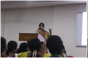
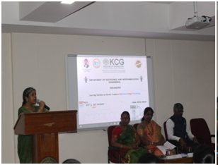
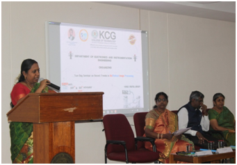
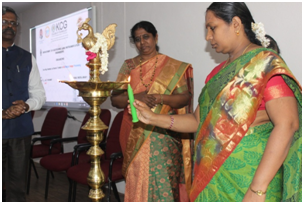
The Department of EIE, KCG College of Technology Conducted Two Days Seminar on “Recent Trends in Biomedical Image Processing” on 29th and 30th November 2017. The Programme started with Inauguration on November 29th 2017. Dr.N.Bharathi , Head of the Department (EIE) and Convener of the Seminar gave the Welcome address. Dr.G.Prabhakaran, Principal, and Dr. Sumathi Poobal, Vice Principal, KCG College of Technology gave felicitation address. Dr.A.K.Jayanthy, Professor-Department of Biomedical Engineering, Kattankulathur Campus, SRM University inaugurated the session and gave inaugural talk. The inaugural session came to an end with the vote of thanks by Ms.Sunetra Banerjee.
(i). “Biomedical Optical Imaging” by Dr. A.K. Jayanthy, Professor, Dept of BME, SRM University, Katankulathur Campus
Dr. A.K Jayanthy started the first session with a brief introduction about Fluorescent Imaging techniques and Laser Speckle Contrast Imaging (LSCI) . Later she gave a detailed lecture on each topic like light sources, advantages of the techniques and its application in LSCI. She presented her case study on Psoriasis and Diabetic ulcers. She showed how optical imaging helped in diagniostic accuracy and specificity when compared to other imaging techniques with clear examples and its results. She also made a note that still optical imaging though promising diagnostic tool has very few people taken up their research in this area and she welcomed aspirants to pursue their research in optical imaging.
(ii) “MRI Brain Image Analysis” by Dr.M.Kayalvizhi, HOD- BME Dpt, Agni College of Technology,Thalambur.
Dr.M.Kayalvizhi started session-II with the basics of MRI brain imaging techniques. She explained the two types of MRI images namely T1 weighted images and T2 weighted images and its anatomical planes. She also explained about the need of skull stripping, various trigonometric parameters use in
the processing of MRI images and its algorithms. Finally the session ended up with the case study of various MRI brain images and its image processing algorithm results using K-Means clustering.
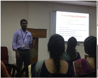
(iii). “Medical Image Analysis Using MATLAB” by Mr.J.Saminathan, Teaching Fellow, Dep. Of ECE, College of Engineering, Guindy
Mr.J.Saminathan started session-III with a brief introduction to various imaging modalities. He then explained various image processing techniques like image acquisition, image pre-processing, image segmentation, image enhancement, feature extraction and classification in MATLAB with various examples. He also explained why we go for gray scale images for image processing when compared to RGB image. He finally demonstrated all the above said techniques with the case study of detection of Lung Cancer using CT images.
(iv). “Imaging Science- An Overview” by Prof. S. Panneer Selvam , Associate Professor Of Medical Physics & RSO,Radiology and Imaging Sciences, Sri Ramachandra Medical College.
The second day of the seminar started with a talk by Professor Panneer Selvam. He started the session with the basics of imaging. He explained the science and Instrumentation behind all the imaging modalities like X-Ray, sonogram, CT, MRI, PET and Nuclear Imaging. He also made the audience understand how the image processing techniques help in improving the image quality in computer aided diagnostics. He also emphasized on the research avenues in this area in India and abroad.
(v) “Recent Advances in Diagnostic Radiology” by Dr. Balaji Jeevanandam, Asst.Professor, Radiology and Imaging Sciences, Sri Ramachandra Medical College.
Dr.Balaji Jeevanandam presented his talk on Recent Advances in Diagnostic Radiology. He shared his experience and views on various techniques in 3D image reconstruction like surface rendering and volume rendering. He explained with various sample images that how 3D image processing techniques help doctors in diagnosing abnormalities in certain crucial cases which 2D image processing technique could not accomplish.
(vi) “Thermography – A Promising Diagnostic Tool” by Dr.Sheeja.V.Franics Associate Professor, Department of ECE,MNM Jain College.
Dr. Sheeja.V.Francis focused on Thermography. She started the session with the basics of thermal imaging and its need as a diagnostic tool. She explained the role of thermal imaging in breast cancer diagnostics. She created awareness that though Mammogram is the conventional diagnostic tool for breast cancer and considered as the golden standard could not help in early diagnosis of breast cancer and also the painful procedure that the patients have to undergo even for screening purposes. Thermal imaging and its processing techniques on the other side though not accepted as a golden standard could help in early diagnosis. She clearly explained the above said point with images and results.
The Seminar came to an end with delivery of certificates to the participants and Valedictory.